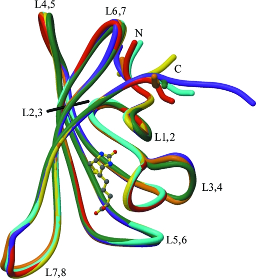
Figure 2.
Polypeptide tracing for the SWT and SWTB subunits after superposition on subunit A of SWT. Subunits colored as in Fig. 1 ▶. The loops are labeled, as are the N- and C-termini. Biotin bound to the A subunit of SWTB is shown in ball-and-stick representation. This figure was drawn with MolScript (Kraulis, 1991 ▶) and RASTER3D (Merritt & Bacon, 1997 ▶).

