Figure 3.

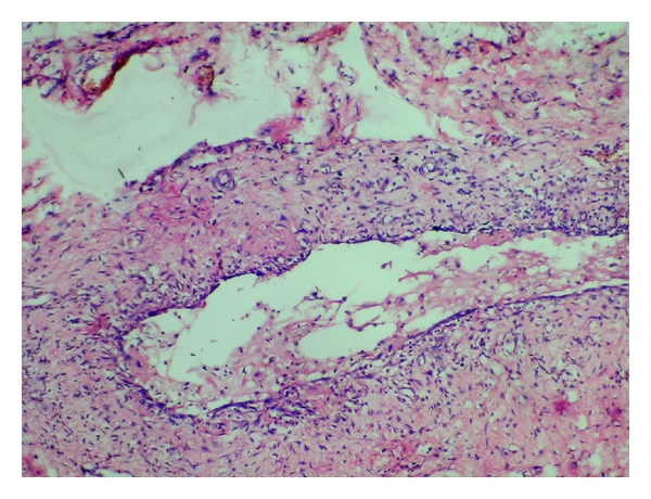
Histopathological picture of the excised lesion showing mucin collection in the lumen lined by connective tissue with inflammatory cells.

Histopathological picture of the excised lesion showing mucin collection in the lumen lined by connective tissue with inflammatory cells.