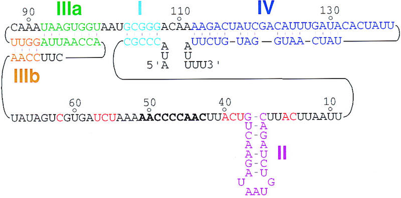
Figure 1.

Telomerase RNA in T. thermophila. The sequence and structure of the T. thermophila telomerase RNA are shown as described previously (Greider and Blackburn 1989; ten Dam et al. 1991; Romero and Blackburn 1991; Zaug and Cech 1995). The representation here emphasizes the secondary structure elements and the primary sequences examined in this study. Stem I is in light blue, stem–loop II is in purple, the stem of stem–loop III as initially described is in green (IIIa), with pseudoknot formation producing a second stem in brown (IIIb), and stem-loop IV is in dark blue. The template region is in bold, and phylogenetically conserved sequences in the single-stranded, template-adjacent regions are in red. RNA 5′ and 3′ ends are indicated.
