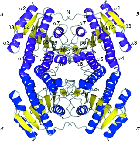
Figure 2.
Crystal structure of R. prowazekii 3-ketoacyl-(ACP) reductase (FabG). FabG from R. prowazekii crystallizes with two molecules per asymmetric unit, which are shown as ribbon diagrams with purple helices and yellow strands. The active-site residues (Ser143, Tyr156 and Arg160) are grouped near the loop connecting α5 and β5. Secondary-structure elements are labeled from both chains. A tetramer is generated by crystallographic symmetry. Molecules A′ and B′ are shown as ribbon diagrams with blue helices and yellow strands. The figure was generated with CCP4mg (McNicholas et al., 2011 ▶).

