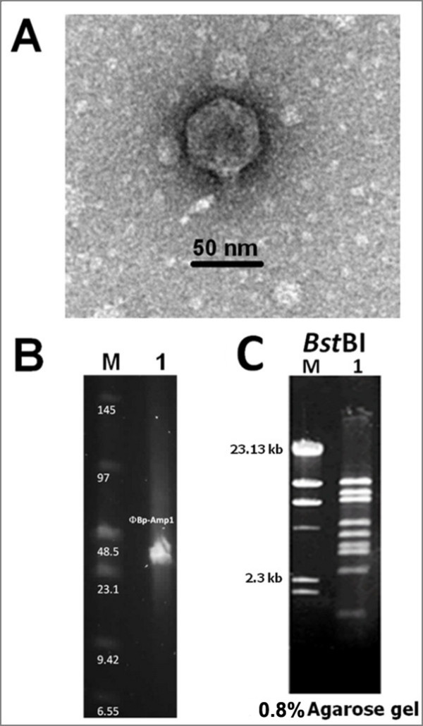
Figure 1.

Electron microscopic and DNA analysis of ФBp-AMP1. (A) Transmission electron micrograph and ΦBp-AMP1 (B) The genome size of ΦBp-AMP1 determined by PFGE. (C) Restriction DNA pattern of ΦBp-AMP1 genomic DNA digested with BstBI (lane1). Lane M, λ HindIII DNA marker.
