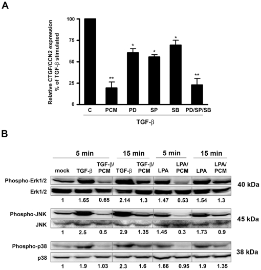
Figure 2. T. cruzi PCM abrogates MAP kinase activation and decreased MAP kinase signaling results in inhibition of ctgf/ccn2 expression.
(A) HFF were stimulated with 5 ng/ml TGF-ß1 in the presence of medium, T. cruzi PCM or with 10 µM SP600125, 20 µM SB203580 or 50 µM PD98059 as indicated for 2 hours prior to mRNA harvest for quantitative real-time PCR analysis. MAP kinase inhibitor treatments were carried out following a 30-minute pre-incubation step. Data is represented as the mean ± s.e. from triplicate experiments (n = 4). Statistical significance was assessed using the Student's t-test (** p<0.01, * p<0.05). (B) Western blot of phospho-Erk, phospho-JNK and phospho-p38 normalized to total Erk, JNK and p38 respectively in lysates from HFF stimulated with 5 ng/ml TGF-ß1 or 10 µM LPA for 5 or 15 minutes in serum-free media or parasite-conditioned medium. Results for densitometric analysis are shown as numerical values below each panel and represented as phosphorylation relative to mock-treated controls (arbitrarily set to a value of 1.0).

