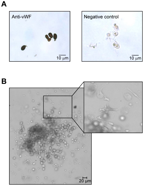
Figure 2. Isolation and characterization of CD133+/VEGFR2+ cells.
Cells sorted by FACS were further characterized by the expression of a specific endothelial cell marker or cultured in a human methylcellulose base media (A and B, respectively). A) CD133+/VEGFR2+ cells were immunohistochemically stained with an antibody against von Willebrand Factor (vWF). Negative control: omission of the primary antibody. B) Phenotypically, colonies formed by these cells in methylcellulose base media show the typical shape of early EPC-colonies with round immature cells in the center and dendritic or spindle cell-shaped peripheral cells (see magnification).

