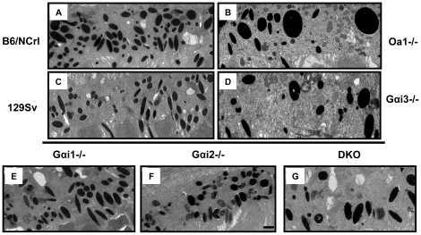
Figure 1. Appearance of RPE melanosomes from Gαi1-/-, Gαi2-/- and DKO Gαi1-/-, Gαi3-/- mice.
RPEs from Oa1-/- (B) and their control B6/NCrl (A) mice and Gαi3-/- (D) and their control 129 Sv (C) mice have been published before [12] and are shown for comparison. The ultrathin RPE sections show that Gαi1-/- (E) and Gαi2 -/- (F) mice do not have macromelanosomes as those present in Oa1-/- (B), Gαi3-/- (D) and DKO Gαi1-/-, Gαi3-/- (G) mice. Electron micrographs of ultrathin sections, 16,000× magnification; scale bars for all micrographs, 01 µm, is shown only in (F).

