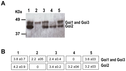
Figure 6. Western blot of RPE proteins from all Gαi-/- mice using an anti- Gαcommon antibody.
A) RPE membrane proteins from 3-month-old mice were separated by SDS-PAGE, blotted and reacted with Gαcommon antibodies. Protein bands were visualized using the ECL detection reagent. The RPE proteins are from: Gαi1-/- (lane 1), Gαi2-/- (lane 2), Gαi3-/- (lane 3), DKO (lane 4) and 129 Sv (lane 5) mice. The membrane was then stripped and reacted with α-tubulin antibodies to confirm that the amount of protein loaded on each lane was the same. B) Analysis of the immunoblot by densitometry using the Quantity One 1-D Software. Numbers represent the optical density units per mm2 (OD U/mm2) of each Gαi protein band.

