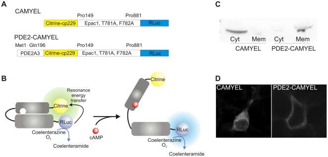
Figure 1. Schematic overview of BRET sensors.
(A) CAMYEL is comprised of cytosolic, catalytic inactive Epac1 sandwiched between Citrine-cp229 and the Renilla luciferase (RLuc). PDE2-CAMYEL is targeted to the membrane by fusion of the N-terminal part of PDE2A3 to CAMYEL. (B) Binding of cAMP to CAMYEL/PDE2-CAMYEL induces a conformational change in the Epac1 part resulting in a decrease of energy transfer from the luciferase to Citrine. (C and D) Western blot and confocal images showing the distribution of CAMYEL in the cytosol and the targeting of PDE2-CAMYEL to the membrane. Cyt, cytosolic fraction; Mem, membrane fraction.

