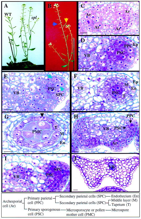
Figure 1.

Wild-type and spl mutant phenotype during anther development. The box at bottom shows wild-type cell lineage and nomenclature. (A) Photograph showing wild-type (WT) and the spl mutant plants. (B) Photograph of a mutant plant in which sterile siliques (blue arrow) are reverted to wild type (yellow arrow), demonstrating the excision of Ds from the spl gene. (C–G) Micrographs of 5 μm-thick cross sections of wild-type anthers at different stages. Stages are according to Sanders et al. (1999). (C) Four archesporial cells (Ar) are visible at the four corners of a stage 2 anther. (D) One developing locule at stage 3 showing the PSC and the PPC. The cells close to the vascular bundle (VB) also divide and participate in the formation of the archesporium. (E) Part of an anther at stage 4 showing the SPCs. (Arrows) Cell wall between newly formed SPCs. (F) Part of an anther at stage 5 showing the formation of tapetum (T), the middle layer (M) and endothecium (En). (G) An anther at stage 6 shows one of the four microsporangia. The microsporocyte or PMC is enlarged just prior to entering meiosis. The tapetum becomes binucleated (arrowhead). (H–J) Micrographs of 5 μm-thick cross sections from spl mutant anthers. (H) An Ar is visible at the lower right corner and PSCs and PPCs at the upper right corner in stage 2 and 3 anthers. (I) Part of an anther at stage 4 showing development of the SPCs. (J) A cross section of a spl mutant anther at maturity. All cells are vacuolated. (Ep) epidermis. Bar, 10 μm.
