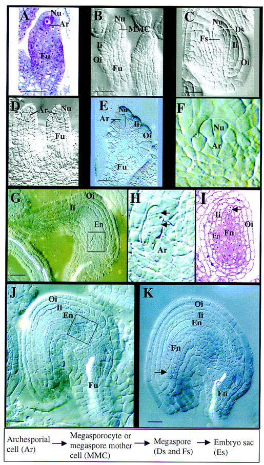
Figure 2.

spl mutant phenotype during ovule development. A–C are from wild-type carpels; D–K are from spl mutant carpels. All except A and I are micrographs of ovules cleared with Herr’s solution viewed under Nomarski optics. Stages are according to Schneitz et al. (1995). The box at bottom shows wild-type cell lineage and nomenclature. (A) Longitudinal section of an ovule primordium at stage 1-II. Note the archesporial cell (Ar) at top. (B) An ovule primordium at stage 2-III showing the megasporocyte (MMC) and the initiation of both inner (Ii) and outer (Oi) integuments. Note the large megasporocyte spanning about three nucellar cells (Nu) longitudinally. (C) An ovule at stage 2-V shows the functional megaspore (Fs) and two degenerating megaspores (Ds) at the top of the ovule. Another degenerating megaspore is not visible on this plane because of the T-shaped configuration. Both Ii and Oi have elongated along the axis of the ovule. (D) Ovule primordia from a mutant carpel showing the presence of archesporial cells (Ar). (E) Mutant ovule at a similar stage to B. Note that the Ar and the nucellus do not elongate longitudinally. The Ii and Oi primordia were initiated. The dotted vertical line delineates the cell wall between the Ar and nucellar cells. (F) A magnification of the boxed area in G shows the arrested Nu and Ar. Note the size of the apical nucellar and the archesporial cell. (G) A mutant ovule shows the development of the Ii and Oi and the differentiation of endothelium (En). No embryo sac is formed and the nucellus is arrested (boxed area). (H) Magnification of the boxed area in J shows the transversal division (arrows) of the apical nucellar cell. Note the size of Ar. (I) An oblique section of an ovule at the same stage as K shows the finger-like nucellar structure (Fn) and the integuments. (Arrow) Transversal division of the apical nucellar cell. (J) Mutant ovule from stage 13 flower showing the initiation of the finger-like nucellar structure (boxed area). (K) Mutant ovule from stage 14 flower showing the finger-like structure (Fn). (En) Endothelium; (Fu) funiculus. Bar, 5 μm.
