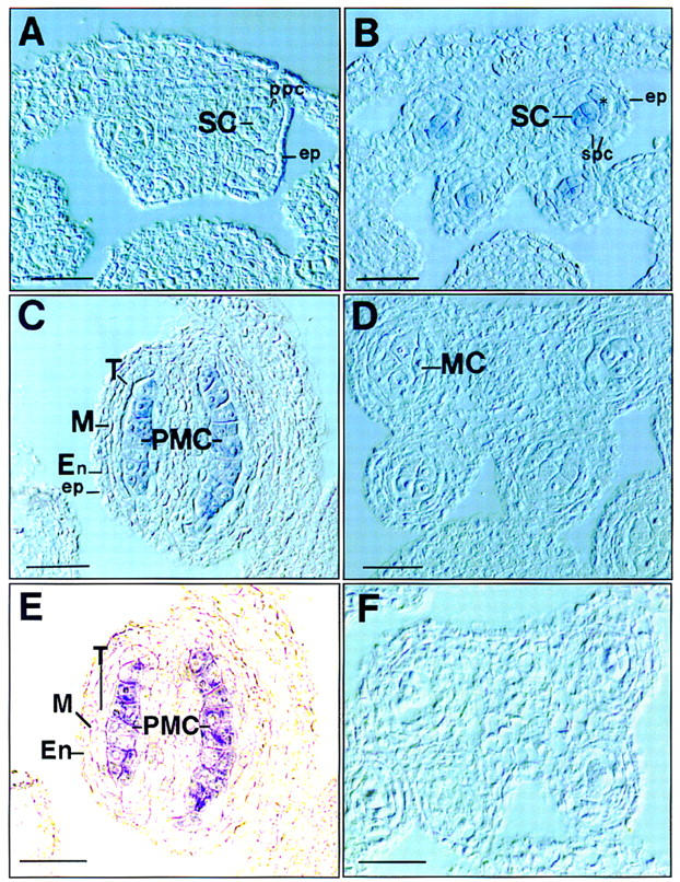
Figure 6.

In situ localization of SPL mRNA in anthers. Sections of anthers at different stages were hybridized with DIG-labeled antisense SPL RNA probe and directly photographed with Nomarski or phase-contrast microscopy without counterstaining. Stages are numbered as described by Sanders et al. (1999). (A) Cross section of an early stage 3 anther shows no hybridization signal. (B) Cross section of an anther at early stage 4 shows SPL expression in sporogenous cells (SCs). (*) newly divided SPCs. (C) Longitudinal section of a stage 5 anther shows SPL expression in microsporocytes or PMCs. (D) Cross section of an anther at meiosis at stage 6 shows no signals in meiotic cells (MCs). (E). Phase-contrast micrograph of the same section as C to highlight the hybridization signals. (F) Cross section of a stage 5 anther hybridized with sense RNA probe as control. (En) Endothecium; (ep) epidermis; (M) middle layer; (ppc) primary parietal cell; (T) tapetum. Bar, 10 μm.
