
Figure 7.
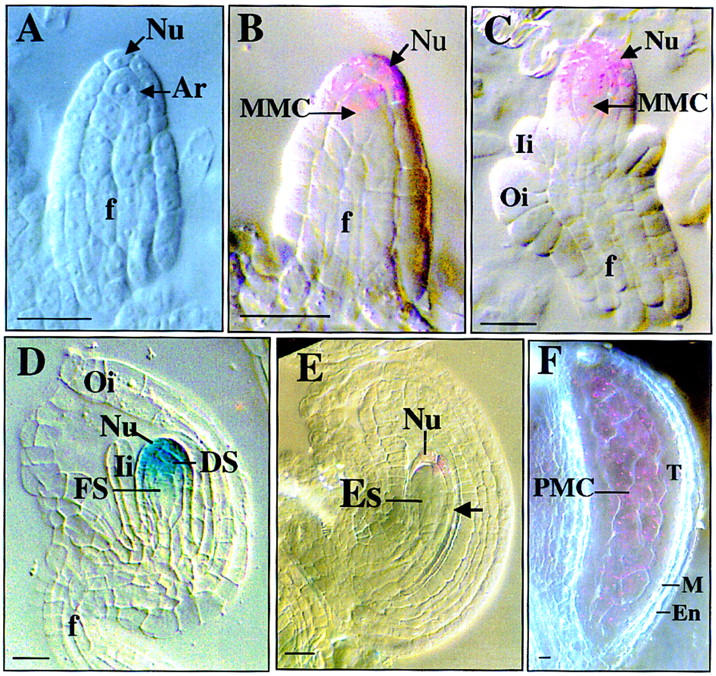
SPL promoter activity monitored by GUS activity.
SPL promoter activities visualized by GUS staining at different stages of ovule development and photographed with Nomarski optics. Each purple staining spot represents an individual indigo precipitate viewed through Nomarski optics due to low level of GUS activity and blue staining as in D is due to strong staining that blocks the polarized light. Stages are according to Schneitz et al. (1995) for sporogenesis and Christensen et al. (1997) for gametogenesis respectively. (A) Ovule primordium at stage 1-II shows no GUS staining in archesporial cell (Ar) and nucellus (Nu). (B) Ovule at stage 2-I showing GUS staining in young MMC and distal nucellar cells. (C) Ovule at stage 2-III shows GUS staining in mature MMC and distal nucellus. (D) Ovule at early FG1 stage showing GUS expression in degenerating nonfunctional megaspores (DS) and distal Nu cells. (E) Ovule at stage FG2 shows weak GUS staining in the distal nucellar cells. Note the absence of staining in the proximal nucellar cells. (F) Micrograph of a stage 5 anther showing GUS expression in PMCs. (En) endothecium; (Es) embryo sac; (f) funiculus; (FS) functional megaspore; (Ii) inner integument; (M) middle layer; (MMC) megaspore mother cell; (Nu) nucellar cell; (Oi) outer integument; (PMC) pollen mother cell; (T) tapetum. Bar, 5 μm.