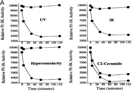
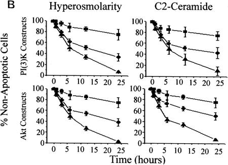
Figure 3.
(A) Stress-induced inhibition of PI(3)K is mediated by acid–sphingomyelinase. MS-1418 NP acid–sphingomyelinase-deficient EBV-transformed (▪) and JY wild-type EBV-transformed human lymphoblasts (•) were treated with C2 ceramide (10 μm), sorbitol (250 mm), IR (10 Gy), or UV-C (10 Jm−2) for the indicated times, and assayed for PI(3)K activity as in Fig. 1B. (B) In cells treated with C2 ceramide or hyperosmolar stress, dominant negative PI(3)K or Akt increases the kinetics of apoptosis and constitutively active PI(3)K or Akt promotes survival. (Top panels) Rat-1 Myc–ER cells were transfected with either an empty vector (•), a constitutively active (p110–SH2) (▪) or a dominant-negative (p85Δ) (▪) form of PI(3)K in addition to a GFP plasmid for transfection identification. The cells were treated for 60 min with 100 nm 4-HT prior to C2 ceramide (10 μm) or sorbitol (250 mm) addition (•). At 24 hr, the cells expressing the GFP plasmid were assayed for apoptosis by Hoechst 33342 and propidium iodide staining. (Bottom panels) Rat-1 Myc–ER cells were transfected with either an empty vector (•), a constituatively active (Myr–Akt) (▪) or a dominant-negative (ΔAkt) (▴) form of c-Akt in addition to a GFP plasmid for transfectant identification. The cells were treated the same as above. Error bars represent the mean of triplicate cultures from a representative experiment.


