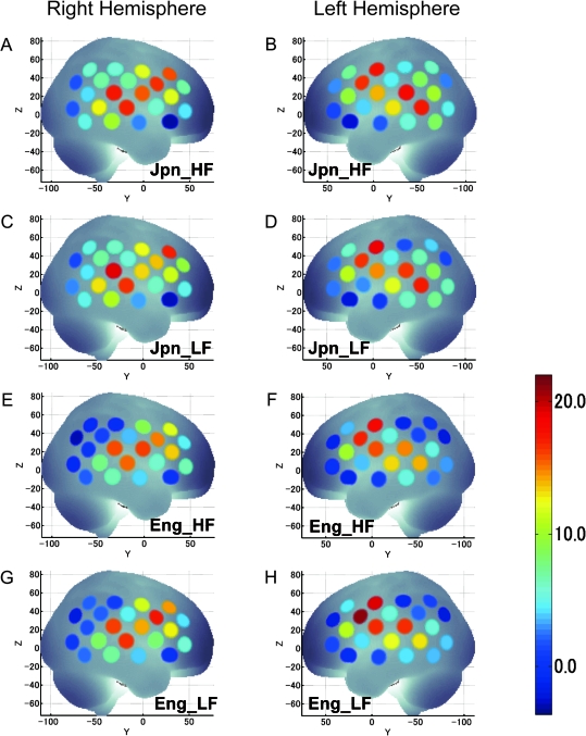
Figure 4.
Cortical activations during word repetition tasks. Average fNIRS data obtained from 392 participants were projected onto the MNI standard brain space by spatial registration. The position of the measurement channels together with cortical activation of both RH and LH are shown in the figures: high-frequency Japanese (Jpn_HF) RH (A) and LH (B); low-frequency Japanese (Jpn_LF) RH (C) and LH (D); high-frequency English (Eng_HF) RH (E) and LH (F); and low-frequency English (Eng_LF) RH (G) and LH (H). The color scale indicates t-values (uncorrected).

