
Fig. 2.
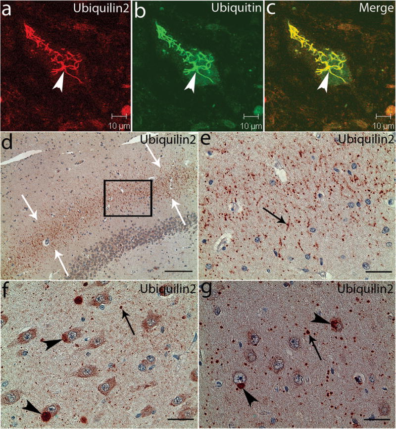
Ubiquilin2-immunoreactive inclusions in the spinal cord and hippocampus. Spinal cord (a–c) and hippocampal (d–g) sections from a patient with a UBQLN2P506T mutation were analyzed with confocal microscopy (a–c) and immunohistochemistry (d–g) using a monoclonal antibody against ubiquilin2 (ubiquilin2-C). The ubiquilin2-positive and skein-like inclusions (arrowhead) are shown in a spinal motor neuron (a). These inclusions are also ubiquitin-positive (b and c). In the hippocampus, the ubiquilin2-positive inclusions are shown in the molecular layer of the fascia dentate (d and e), CA3 (f) and CA1 (g). White arrows in the panel (d) indicate the middle region of the molecular layer with ubiquilin2-positive inclusions. The higher magnification image of the boxed area in panel (d) is shown in panel (e). Black arrows indicate the representative inclusions in neurites (e–g), and arrowheads indicate cytoplasmic inclusions in the cell bodies (f and g). Scale bar, 200μm in panel (d), 50μm in panel (e) and 25 μm in panels (f and g).