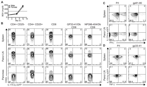
FIGURE 4.
T cells do not produce IL-17A in diabetic RIP-LCMV-GP mice. IL-17A eGFP reporter RIP-LCMV-GP mice were infected with 104 pfu LCMV Armstrong (n=8). A Blood glucose levels of male (black symbols), or female (open symbols) IL-17A eGFP reporter RIP-LCMV-GP mice at indicated time points a.i. B IL-17A/eGFP expression in various T cell types from spleen (upper row), pancreatic LN (middle row), or pancreas (lower row) of diabetic mice (day 14 a.i.). Cells were freshly stained and acquired without prior in vitro restimulation, inhibition of protein secretion, cell fixation or permeabilization. Graphs shown are gated on the cell type indicated on top of the column. Values <0.1 are depicted as zero. C, D Lack of IL-17A expression by restimulated CD4 T cells (C) and CD8 T cells (D) from diabetic RIP-LCMV-GP mice. IL-17A eGFP RIP-LCMV-GP mice were infected with 104 pfu LCMV Armstrong (n=6). At day 14 post infection (recent onset diabetes), spleen (top row) and pancreatic LN (bottom row) were harvested and, to allow IFN-γ/TNF-α visualization, were restimulated in vitro with PMA and ionomycin (P/I), gp33-41 peptide or gp61-80 peptide in the presence of Brefeldin A, as indicated. Representative graphs shown are gated on CD8 or CD4 T cells in panel C or D, respectively.

