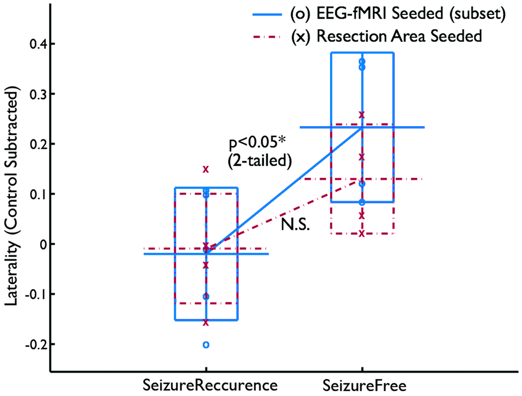
Figure 4.
Comparison between the resection area-seeded functional connectivity analysis and the spike correlated fMRI seeded functional connectivity analysis based on 9 patients. The resection area-seeded functional connectivity analysis did not show a significant difference of laterality between seizure-recurrence and seizure-free groups, whereas the spike correlated fMRI seeded connectivity did. However, the lack of interaction indicates that there was not a significant difference between two seeding methods.

