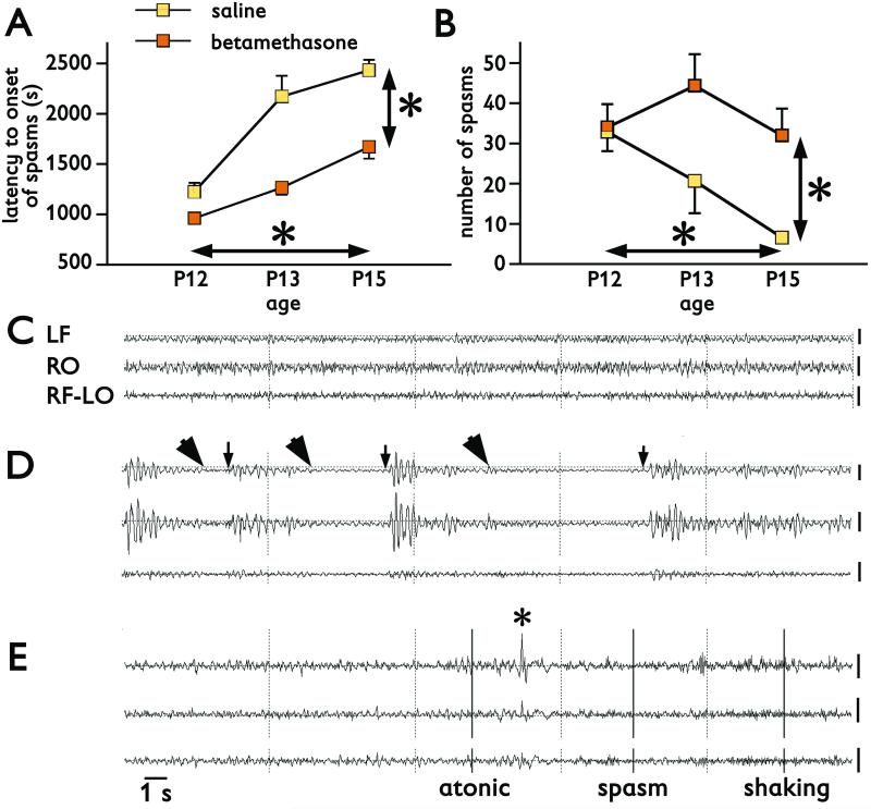
Figure 2. Spasms triggered by repeatedly injected NMDA on P12, P13, and P15 and their EEG correlates.
(A) Latency to onset of spasms triggered by repeated NMDA in P12 (7.5 mg/kg), P13 (12 mg/kg), and P15 (15 mg/kg) rats is significantly shorter in prenatally betamethasone-exposed rats compared to saline controls (vertical *p<0.05). Latency to onset of spasms significantly increases with age (vertical *p<0.05).
(B) Similarly, the number of spasms is significantly increased in prenatally betamethasone-exposed rats compared to saline controls (vertical *p<0.05). There was also a significant decrease in number of spasms with age (horizontal *p<0.05).
(C) EEG recordings in a P13 rat prior to NMDA administration (baseline). LF, RO – left frontal and right occipital channel in monopolar montage versus common reference in the nasal bone. RF-LO – a bipolar montage combined from the right frontal and left occipital channel. Background consists of low-amplitude fast activity.
(D) EEG recording in the same P13 rat after the spasms were triggered with 12 mg/kg of NMDA i.p.. Arrowheads mark onsets of spasms, arrows indicate the end of the spasms. Each of the three spasms was associated with a significant decrease in the EEG amplitude, an electrodecrement. Between the spasms, large-amplitude irregular waves occurred, a possible rat correlate to hypsarrhythmia.
(E) EEG recordings in another P13 rat prior to the second NMDA trigger. This rat exhibited and atonic/astatic episode, with the beginning marked in the EEG with the first bar and ending at the asterisk. Then a spasm occurred associated with a decrement in EEG amplitude followed by long-lasting head shaking (vertical bars at the onset and the legend).
Calibration 200 μV at the end of each trace. Time mark 1 s for all recordings.

