
Figure 2. Vascularization and topographical cues for engineered cardiac tissues.
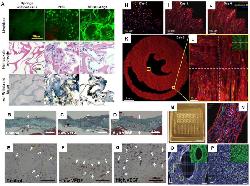
(A) Tube formation after 7-day cultivation of endothelial cells on collagen scaffolds with covalently immobilized vascular endothelial growth factor (VEGF) and angiopoietin-1 (Ang1). Images of PBS control group (scaffold with no growth factor) and VEGF+Ang1 group (scaffold with immobilized VEGF and Ang1) with live/dead staining (CFDA stains live cells green, PI stains dead cells red), hematoxylin and eosin staining, and von Willebrand factor staining. Sponges without cells are also shown. Random clusters of cells were found in PBS control group, compared to circular structures formed by elongated thin cells with nuclei in the peripheral position in the VEGF+Ang1 group ((A) with permission from [34]). (B-G) Implantation of collagen scaffolds with covalently immobilized VEGF to replace a full right ventricular free wall defect in rat hearts (B-D) Masson’s trichrome staining at 28 days after repair of right ventricular free wall defect shows higher patch thickness for patches with low dose VEGF (14.5 ± 1.4 ng VEGF) and high dose VEGF (97.2 ± 8.0 ng VEGF), compared to control patch with no VEGF. Arrows indicate thickness of the patch. (E-G) CD31 staining shows more CD31-positive vascular structures (indicated by arrows) for patch with high dose VEGF, compared to control low dose VEGF patch ((B-G) with permission from [36]). (H-L) Formation of realistic cardiac microstructure using micropatterned anisotropic slice cultures that replicate the natural fiber directions in ventricular cross sections. (H-J) The cells attached, spread and aligned to the micropatterned fibronectin lines and formed confluent cardiac fibers by Day 6. (K) A composite image of the entire micropatterned slice culture as well as (L) close-up images of four sections with the underlying fibronectin pattern (green, inset) is shown ((H-L) with permission from [44*]). (M-P) PDMS molds with arrays of (M) mesoscopic posts were used to fabricate three-dimensional muscle tissue architectures. (N) After 2 weeks of cultivation, tissue constructs were formed with densely aligned and striated cardiomyocytes, showing a high level of connexin-43 expression.. Directions of cell alignment in subregions (blue squares) were obtained to show more aligned cells in (O) PDMS molds with posts compared to (P) those without posts. (P,O) Inset shows the same image with F-actin stain (green) ((M-P) with permission from [47] ).