
Figure 1. Naïve B cell access to the outer follicle is promoted by EBI2.
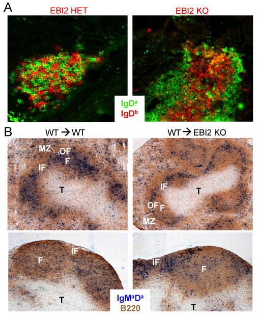
(A) Isolated lymphoid follicles in the small intestine of 50:50 mixed WT or EBI2-/- Ighb (red) and WT Igha (green) BM chimeras, stained as indicated. (B) Spleen and pLN sections from WT or EBI2 deficient mice that had received one day transfers of WT (Igha) B cells. Stained to detect the transferred B cells (IgMaDa, blue) and endogenous B cells (B220, brown). F, follicle (a single follicle is labeled); IF, inter follicular region; OF, outer follicle; T, T zone; MZ, marginal zone.