
Figure 3. EBI2 functions together with CCR7 and CXCR5 to distribute activated B cells along the B-T boundary.
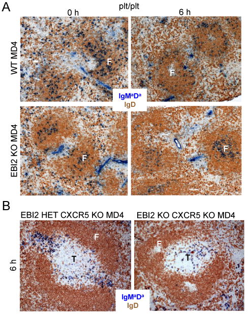
(A) Distribution of WT and EBI2 KO MD4 B cells in CCR7-ligand deficient (plt/plt) recipient spleens at 0 or 6 h after HEL antigen injection. (B) Distribution of EBI2 het CXCR5 KO and EBI2 CXCR5 DKO MD4 B cells in the spleen of WT hosts 6 h after HEL antigen injection. Transferred MD4 B cells were detected by staining for IgMa and IgDa (blue) and endogenous B cells with antibodies to total IgD (brown). Views are representative of three mice.