
Figure 4. CD40 engagement promotes movement of antigen-activated B cells to the outer follicle.
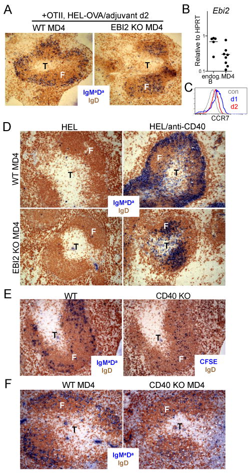
(A) Distribution of activated WT and EBI2 KO MD4 B cells in the spleen at day 2 of the response to HEL-OVA in adjuvant in the presence of OTII helper T cells. (B) EBI2 transcript abundance in sorted day 2 activated WT MD4 B cells (of the type in A) and endogenous B cells. Data are pooled from three experiments. (C) CCR7 surface abundance on activated B cells at day 1 and day 2 of the T-dependent response in mice of the type in A. Data are representative of two experiments. (D) Distribution of WT (upper panels) or EBI2 KO (lower panels) MD4 B cells in the spleen of CD40-deficient hosts, 2 days after immunization with HEL and treatment without or with anti-CD40. (E) Distribution of activated WT and CD40 KO B cells in the spleen at day 2 following co-transfer into bm12 recipients. Serial sections were stained to detect WT (IgMaDa) or CD40 KO (CFSE) transferred B cells (blue) and endogenous B cells (IgD, brown). (F) Distribution of WT and CD40 KO MD4 B cells in the spleen at day 2 of the response to HEL-OVA in adjuvant in the presence of OTII helper T cells. Transferred MD4 B cells in A, D and F were detected by staining for IgMa and IgDa (blue) and endogenous B cells with antibodies to total IgD (brown).