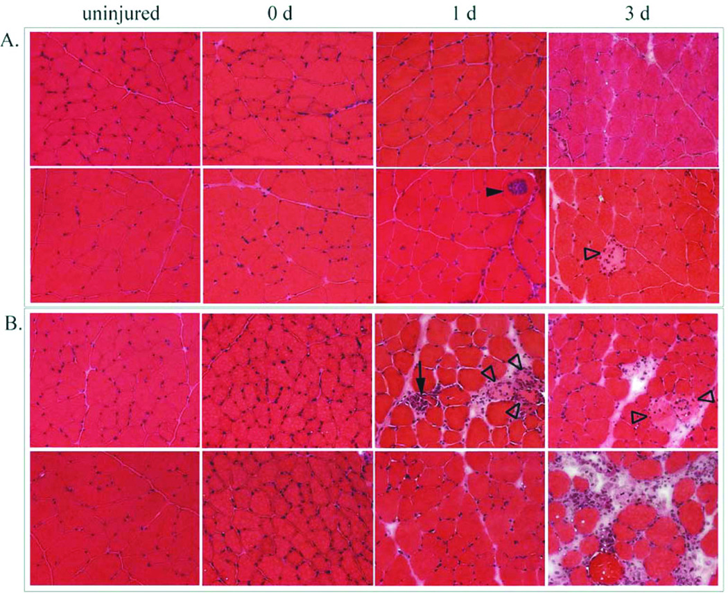
Figure 2.
Time course of injuries, shown in muscle sections stained with hematoxylin and eosin (H&E). Panel A: LSI produced some fibers that were centrally invaded by inflammatory cells (closed arrowhead) and swollen, pale and peripherally invaded fibers (open arrowhead). Panel B: SSI produced local areas of interstitial edema with inflammatory cell accumulation, pale, swollen, and peripherally invaded fibers (open arrowheads) and fully invaded fibers (arrow). The inflammatory response was greater after the SSI.

