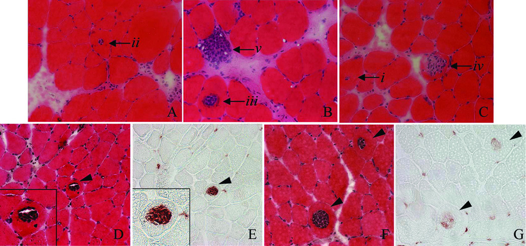
Figure 3.
Central necrosis observed 1 d after LSI. A–C. H&E stained muscles showing the likely progression of centrally invaded fibers (starting with i and progressing to v). D, E: Serial sections of muscle stained with H&E and labeled for ED1-positive macrophages. F, G: Serial sections of muscle stained with H&E and labeled for ED2-positive macrophages. In this example, fibers were centrally invaded primarily by ED1-positive macrophages 1 d after LSI.

