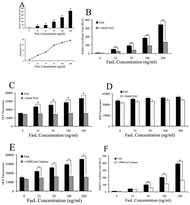
Fig. 1. FasL-dependent NF-κB activity is mediated by hydrogen peroxide.
A. Cells were co-transfected with 100ng/well of NF-κB-Luc and 10ng/well of pRL-tk normalizing luciferase plasmid for 24 h. Transfected cells were treated with increasing concentrations of FasL (0–200ng/ml) for 12 h and assayed for NF-κB luciferase activity. Plots show relative NF-κB activity over non-treated control (*P < 0.05 for each FasL data point as compared to control without FasL). Concurrently, cells that were treated with increasing concentrations of FasL were assayed for apoptosis using the Hoechst assay and graphed. B. Cells co-transfected with the NF-κB-Luc (100ng/well) and pRL-tk vectors (10ng/well) for 24 h. Transfected cells were pre-treated with 10mM NAC for 1 h followed by treatment with increasing doses (0–200ng/ml) of FasL for 12 h. NF-κB activity was measured by luciferase assay. C and D. Cells were either left untreated or were pre-treated with 10mM NAC for 1 h followed by treatment with various concentrations (0–200ng/ml) of FasL. Cells were then analyzed for either (B) H2O2 or (C) ·O2− production by measuring DCF and DHE fluorescence intensity, respectively. Plots show relative fluorescence intensity over non-treated control at the peak response time of 1 h. E. Cells were pre-treated with 1000U/ml catalase for 1 h, followed by FasL treatment (0–200ng/ml) for 12 h and were analyzed for H2O2 production by measuring DCF fluorescence intensity. Plots show relative fluorescence intensity over non-treated control at the peak response time of 1 h. F. Cells co-transfected with the NF-κB-Luc (100ng/well) and pRL-tk vectors (10ng/well) were pretreated with 1000U/ml catalase for 1 h prior to treatment with FasL (0–200ng/ml) for 12 h. FasL-dependent NF-κB activity was measured by luciferase assay.

