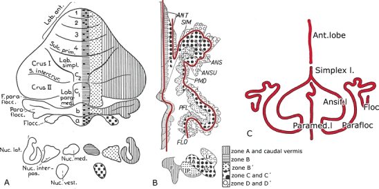
Fig. 4.

A Diagram of the corticonuclear projection of the cerebellum, showing the vermal, intermediate, and lateral zones of Jansen and Brodal [15]. Nomenclature of the lobules according to Bolk [20]. B Diagram of the flattened cerebellar cortex of the cat showing the corticonuclear projection. From Voogd [19]. The contoures (red) lines indicate the direction of the folial chains of vermis and hemisphere. C Stick diagram of the folial chains of the mammalian cerebellum. Bolk [20]. ANS(if.) ansiform lobule, ANSU ansula, ANT anterior lobe, F fastigial nucleus, FLOC flocculus, IA anterior interposed nucleus, IP posterior interposed nucleus, Lc dorsal part lateral nucleus, Lr ventral part lateral nucleus, PFL paraflocculus, PMD paramedian lobule, SIM lobulus simplex
