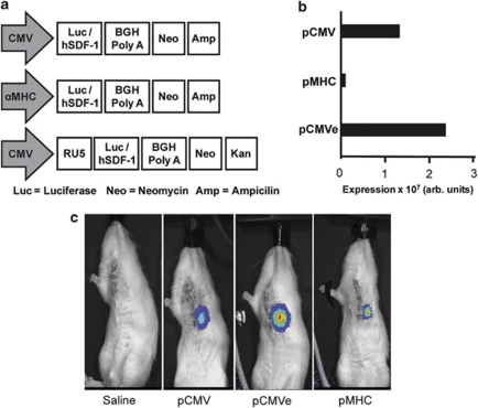
Figure 1.
(a) Schematic of the vector design. Vector design of pCMV, pCMVe and pMHC. (b) HEK293 cells were transfected with pCMV and pCMVe, whereas H9C2 cardiac myoblasts were transfected with pMHC. (c) Cardiac bioluminescence after direct injection. Animals were injected with vectors immediately following LAD ligation and then imaged 3 days after, following an i.p. injection of luciferin.

