
Figure 2.
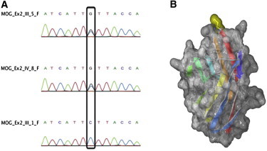
MOG Mutation
Chromatograph example displaying the identified heterozygous mutation (A) in the second exon of MOG (C398G) in two affected (III-5, IV-8) and one healthy family member (III-1). A model of the Ig-like domain of human p.Ser133Cys MOG mutant (B). Secondary structure elements are colored from blue (N terminal) to red (C terminal), and the location of the p.Ser133Cys mutation is highlighted on the molecular surface in yellow, at the top of the image. The image was prepared with Swiss-PdbViewer and rendered with POV-Ray.