
Figure 1.
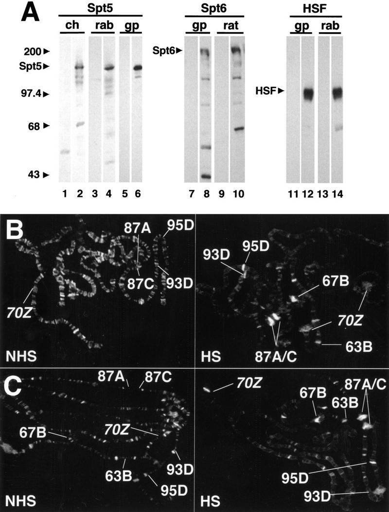
Spt5 and Spt6 are recruited to heat shock puffs on Drosophila polytene chromosomes. (A) Western blot analyses of Spt5, Spt6, and HSF antibodies. Proteins from Drosophila Kc cells were separated on an 8% polyacrylamide gel and transferred to nitrocellulose. Blots were probed with antiserum (even lanes) or with preimmune serum (odd lanes) at a dilution of 1 : 1000. Appropriate secondary antibodies were used and bands visualized using ECL (Amersham). Aligned nitrocellulose strips for each antibody are from the same transferred gels. (Ch) chicken; (rab) rabbit; (gp) guinea pig; (rat) rat. (B,C) Staining of non–heat shock (NHS) and heat shock (HS) polytene chromosomes with rabbit α-Spt5 (B) or guinea pig α-Spt6 (C) antibodies. Major, endogenous heat shock loci (87A, 87C, 63B, 67B, 93D, and 95D) as well as the transgenic sites (hsp70–LacZ [70Z]) are defined. Note the redistribution of Spt5 and Spt6 on heat shock. The staining pattern of both Spt5 and Spt6 is quite similar to that seen previously with cyclin T (Lis et al. 2000).