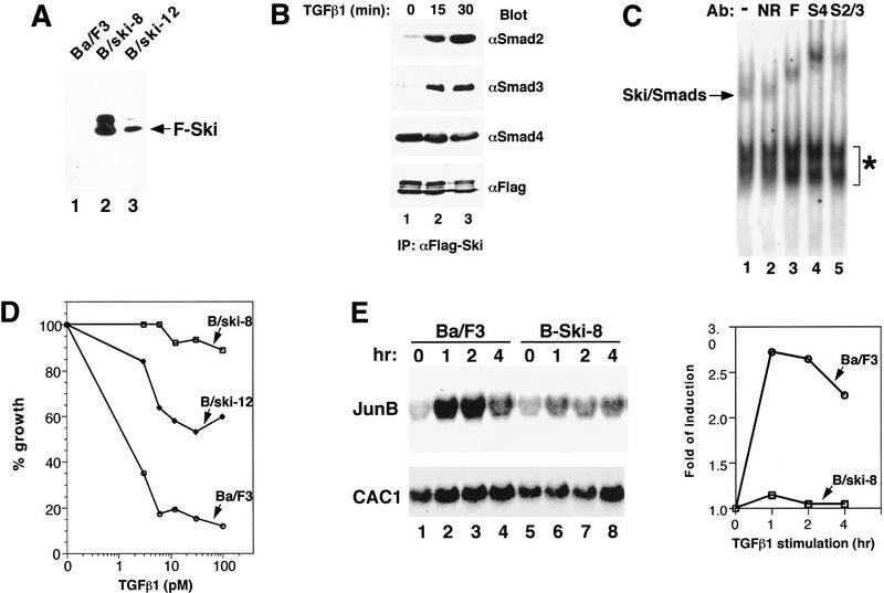
Figure 6.
Overexpression of Ski greatly attenuates TGFβ-induced growth inhibition and activation of JunB expression. (A) Ba/F3 cell lines stably expressing Flag–Ski were generated by retroviral infection. Flag–Ski was isolated by immunoprecipitation with anti-Flag agarose from uninfected Ba/F3 cells or from two stable pools (B/ski-8 and B/ski-12) and analyzed by Western blotting with an anti-Flag mAb. (B) B/ski-8 cells (2 × 10⁸) were stimulated with 200 pm TGFβ1 for 15 or 30 min as indicated. Endogenous Smad proteins associated with Flag–Ski were isolated by immunoprecipitation with anti-Flag agarose and detected by Western blotting with anti-Smad2, anti-Smad3, or anti-Smad4 antibodies. (C) Nuclear extracts were prepared from B/ski-8 cells that had been stimulated with 200 pm TGFβ for 30 min and incubated with 32P-labeled SBE in an EMSA assay. Antibodies used in the supershift assay: (S2/3) anti-Smad2/3; (S4) anti-Smad4; (F) anti-Flag; (NR) nonrelevant control antibody. (*) Non-specific background bands. (D) Growth inhibition assay. Uninfected Ba/F3 cells, B/ski-8 or B/ski-12 cells were incubated for 5 days with various concentrations of TGFβ1 as indicated. The growth of cells was quantified by cell counting and compared to unstimulated cells. The growth rate of B/ski-8 or B/ski-12 cells in the absence of TGFβ1 is similar to that of uninfected Ba/F3 cells. (E) Activation of JunB expression in uninfected Ba/F3 or B/ski-8 cells by Northern blotting. Cells were serum starved for 16 hr and stimulated with 100 pm TGFβ1 for various periods of time as indicated. An analysis of the JunB and human CAC1 RNA is shown. CAC1 was used as a control for equal loading. A quantification of the Northern blot was carried out using the Bio-Rad Molecular Imager FX system and folds of induction of JunB expression are shown in the graph.

