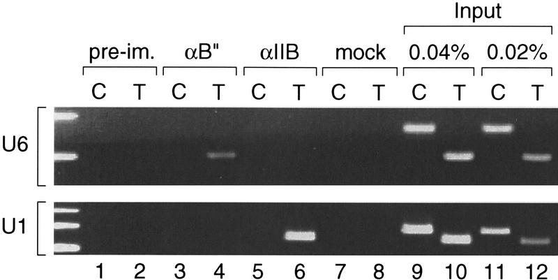
Figure 5.
hB′′ is found in the U6 promoter region in vivo. Rapidly growing HeLa cells were treated with formaldehyde; then cross-linked chromatin was extracted, sonicated, and used as starting material for immunoprecipitations with beads coated with either preimmune (lanes 1 and 2), anti-hB′′ (lanes 3 and 4), or anti-TFIIB (lanes 5 and 6) antibodies, or just beads alone (lanes 7 and 8). The immunoprecipitated material was then analyzed by PCR with test (T) primers specific for the U6 (upper panel) or U1 (lower panel) promoters, or control (C) primers hybridizing to a unique region located 4 kb upstream of the human U6 snRNA gene (upper panel) or a unique region within the 45-kb U1 repeat (Bernstein et al. 1985) 7 kb upstream of the U1 gene (see Materials and Methods for details). Lanes 9–12 show PCRs performed with the test (T) or control (C) primers with 0.04% and 0.02% of the input material used for immunoprecipitation, as indicated above the lanes.

