
Figure 2.
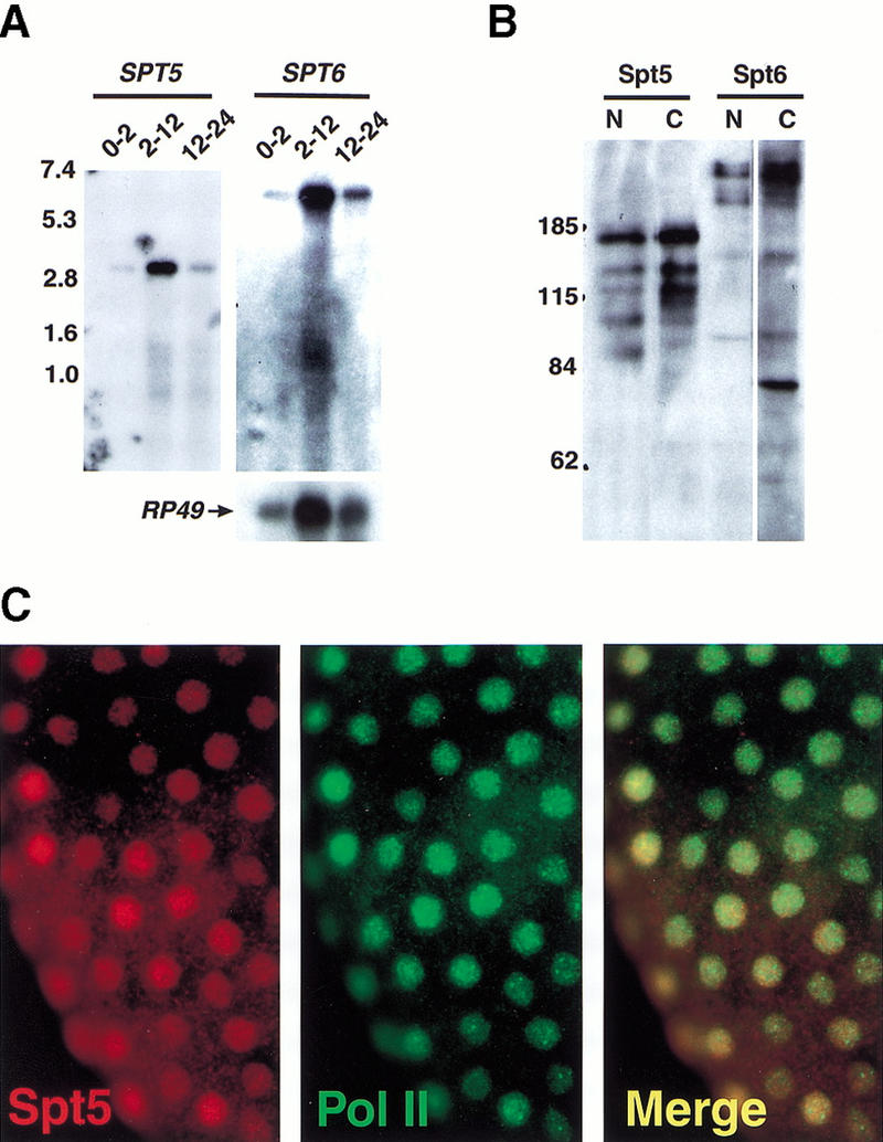
Detection of Spt5 and Spt6 mRNA and protein in Drosophila embryos. (A) Northern analysis of Spt5 and Spt6 transcripts in Drosophila embryos using Poly(A)+ RNA isolated from Drosophila embryos at specific times (indicated in hours) after egg deposition (1.5 μg RNA/lane). The lower panel shows the control probe RP49. Numbers to the left indicate sizes (kb) of molecular weight markers. (B) Western analysis of Spt5 and Spt6. Each lane contains 200 μg of Drosophila 2–12 h embryonic extract. Lanes were then separated and probed with antibodies directed against Spt5, Spt6, or Cyclin T. Antibodies used: lane 1, HMGP11 (directed against amino terminus Spt5); lane 2, HMGP14 (carboxyl terminus Spt5); lane 3, HMGP15 (directed against amino terminus Spt6); lane 4, HMGP17 (carboxyl terminus Spt6). Numbers to the left indicate sizes (kD) of molecular weight markers. (C) Whole mount immunofluorescence detection of Spt5 in Drosophila embryos. A precellularization embryo probed with HMGP11 (left panel); or with an antibody directed against the Pol II CTD as a nuclear marker (middle panel); (right panel) overlay of left and middle panels showing colocalization of red and green signals (appears yellow). All Spt5 and Spt6 antibodies give the same ubiquitous, nuclear staining pattern in both early- and later-staged embryos (data not shown).