
Figure 1.
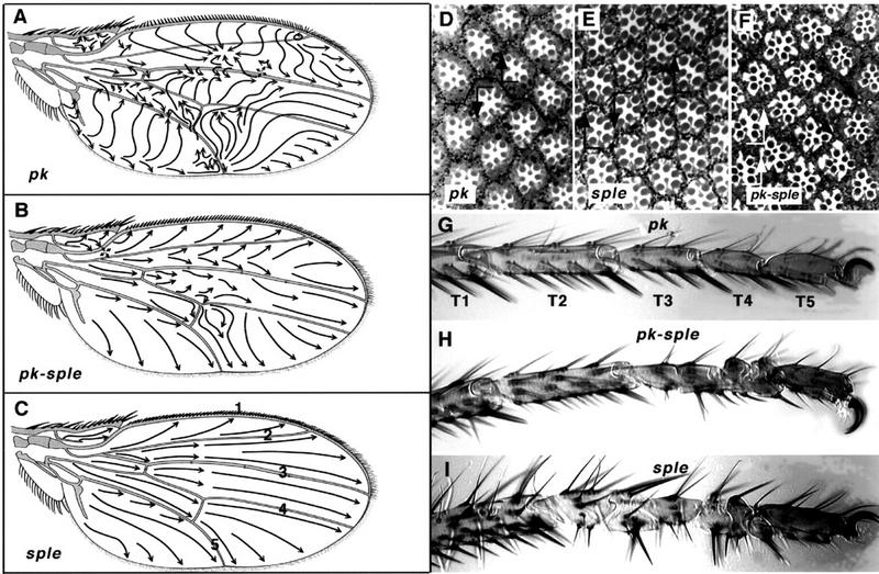
Pk mutant phenotypes. Complete lack of Pk function, in pkpk–sple alleles, gives a weak polarity phenotype in the wing, notum, abdomen, eye, and legs. pkpk alleles cause an extreme polarity phenotype in the wing and notum; pksple alleles affect eye, abdomen, and leg. The pkpk wing phenotype shows a characteristic reversal in the triple-row bristles along the anterior margin, a whorl in the wing hairs near the tip of vein 2, and abrupt discontinuities in hair polarity, e.g., pkpk1 (A). The weak PkPk–sple phenotype shows a slight effect on triple-row bristle orientation and gives gently curved hair polarity vectors, e.g., pkpk–sple13 (B); sple alleles are completely wild type, e.g., pksple1 (C). Arrows indicate the direction along which hairs are aligned; wing veins are designated 1–5 (C). The eye phenotype is wild-type in pkpk1 (D), showing a line of mirror symmetry along the equator (line). On both sides of the equator the R3 photoreceptor cell is aligned towards the pole, in the direction of the arrowhead. In addition to being rotated through 180° ommatidia show reversed chirality around the equator, so that both a rotation and a reflection in the plane of the epithelium is required to superimpose the ommatidial patterns. (E) pksple1 eyes contain a mixture of ommatidia with reversed polarity and chirality in both hemispheres of the eye. These ommatidia remain aligned along the polar axis, but with their R3 photoreceptors directed toward the equator rather than the pole giving rise to D/V mirror-image reversals of the normal rhabdomere pattern. In addition, all the pksple alleles show out ∼1% anteroposterior (A/P) reversed ommatidia (not shown). (F) pkpk–sple13 eyes contain a mixture of chiral forms of ommatidia. Some ommatidia fail to rotate properly, and the resulting imperfections in the hexagonal stacking give a slightly rough eye phenotype. Some ommatidia are aligned at 60° to the equator (black arrows) and some show A/P reversals (white arrows), with the R3 rhabdomere anterior to R4. (G) The tarsi of pkpk1 are wild type; tarsal segments are numbered T1–T5. (H) In pkpk–sple13, the T3 and T4 segments carry medial duplications of the proximal and distal joint structures, with the middle of each segment deleted. This results in alternating reversed-proximal and reversed-distal tarsal joint structures with half the length of a normal segment. (I) In pksple1 the tarsal duplications affect T2, T3, and T4 segments, with an occasional incipient ectopic joint in the distal T1. The distal T5 segment remains unaffected in all mutant alleles.