
Figure 5.
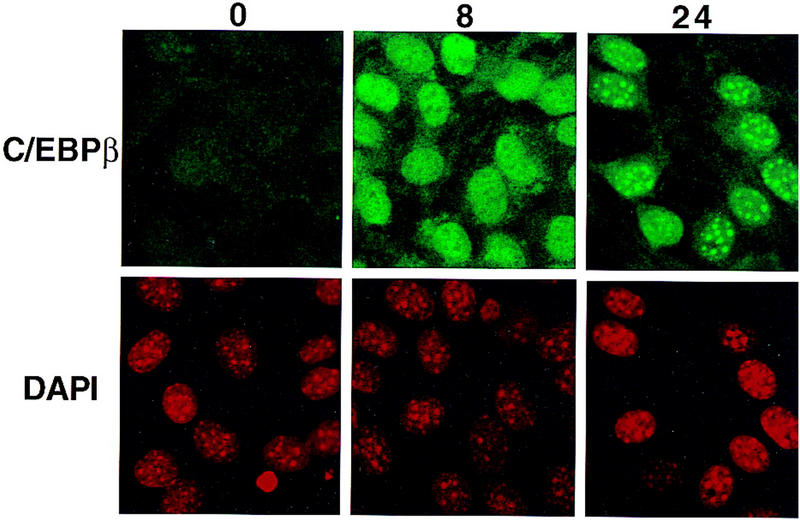
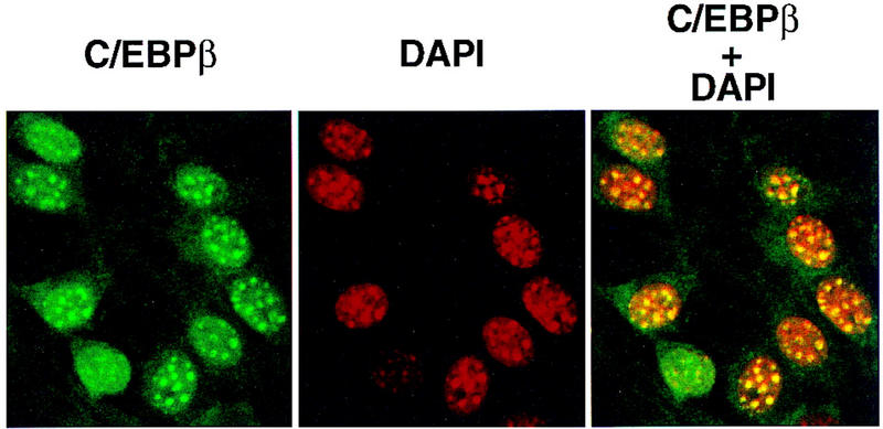
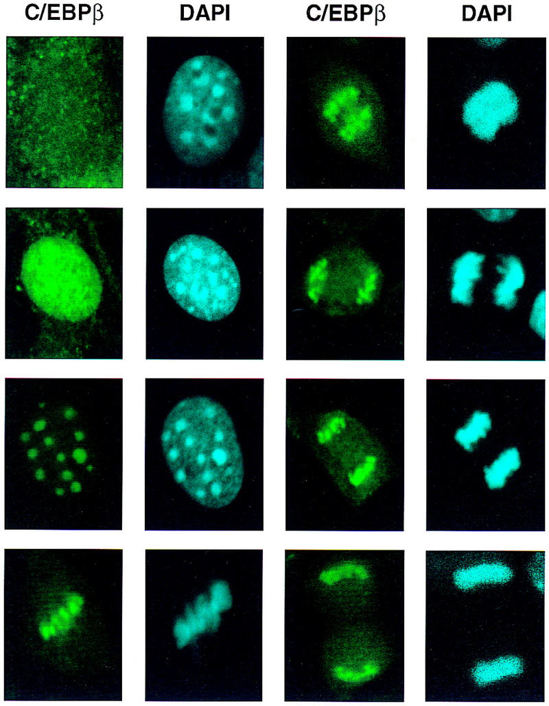
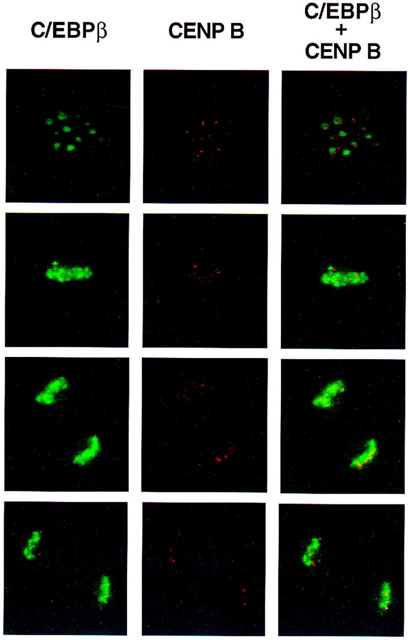
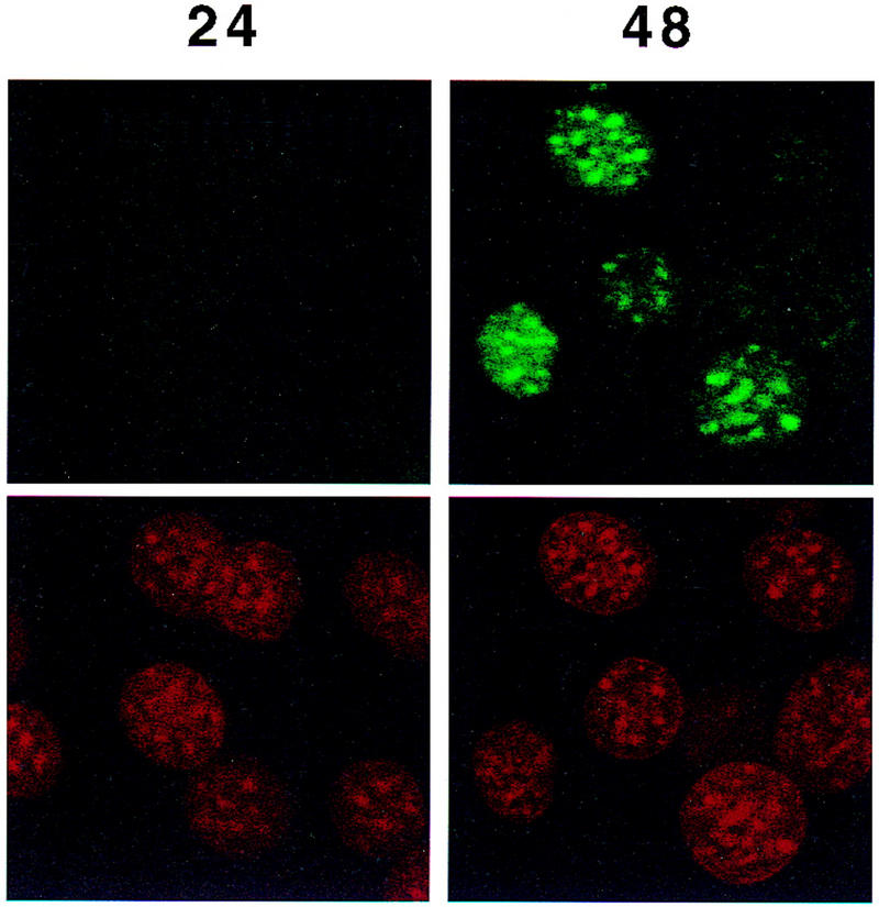
Colocalization of C/EBPβ, C/EBPα, and CENP-B and DAPI staining during differentiation of 3T3-L1 preadipocytes. 3T3-L1 preadipocytes were induced to differentiate using the standard protocol, fixed, treated with antibodies (and FITC-labeled anti-rabbit IgG), and DAPI at the times indicated. Fluorescence images were obtained by confocal microscopy. (A, top) Immunofluorescence with antibody against C/EBPβ and FITC-labeled anti-rabbit IgG. Similar results were obtained with antibody against C/EBPδ (results not shown). (Bottom) Immunofluorescence imaging of the same field as in A with DAPI. (B) Dual fluorescence imaging of C/EBPβ and DAPI by confocal microscopy of cells 24 hr after induction of differentiation. (C) Fluorescence imaging of 3T3-L1 preadipocytes treated with C/EBPβ antibody and DAPI during the first round of mitotic clonal expansion. These selected images of preadipocytes at 28 hr after induction of differentiation represent various stages of the cell cycle. (D) Fluorescence imaging of 3T3-L1 preadipocytes treated with antibodies against C/EBPβ and CENP-B during the first round of mitotic clonal expansion. These selected images of preadipocytes at 28 hr after induction of differentiation represent various stages of the cell cycle. The column on the right represents dual fluorescence images of the two columns of images to the left. (E) Colocalization of immunofluorescence of C/EBPα antibody and DAPI in the terminal stages of the differentiation program.