
Figure 6.
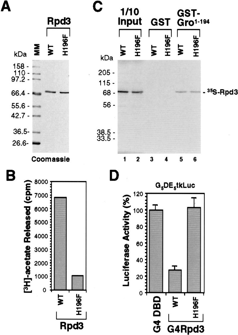
Functional analysis of a single point mutation of Rpd3. (A) A Coomassie blue-stained SDS–polyacrylamide gel showing equal amounts of purified wild-type (WT) and mutant forms of Rpd3. In mutant Rpd3, a conserved histidine residue (H196) has been changed to phenylalanine (H196F). (B) In vitro histone deacetylase assays of purified wild-type (WT) and mutant (H196F) forms of Rpd3. (C) GST pull-down assays show that wild-type (WT) and mutant (H196F) forms of Rpd3 bind to Gro with comparable affinities. GST-pull down assays are conducted as described in Fig. 4D. (D) Gal4 fused to wild-type Rpd3 (WT) is able to repress transcription in S2 cells, whereas Gal4 fused to the single-point mutant form of Rpd3 (H196F) is not able to repress transcription in S2 cells. Cotransfection assays were conducted as described in Fig. 5C.