Figure 7.
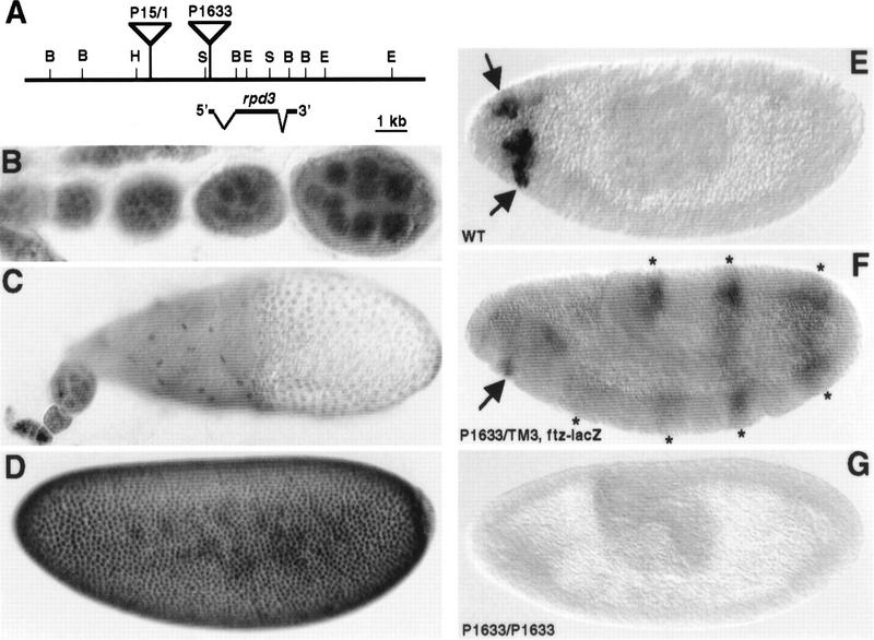
Spatial expression pattern of rpd3 during Drosophila oogenesis and embryogenesis. (A) Genomic organization of the rpd3 gene. The map of the rpd3 transcription unit (at cytological map location 64C1-2) is based on previous reports (DeRubertis et al. 1996; Maixner et al. 1998). BamHI (B), EcoRI (E), HindIII (H), and SalI (S) sites are indicated. The P1633 and P15/1 P-element insertion sites are depicted by inverted triangles. (B–E) Staining of wild-type ovaries and embryos with anti-Rpd3 antibodies. (B) Ubiquitous germ-line nuclear expression of Rpd3 is observed in the wild-type ovary during early oogenesis (before stage 8). (C) Uniform expression of Rpd3 is observed in follicle cell nuclei by stage 10 of oogenesis. (D) Rpd3 is uniformly distributed throughout the nuclei of precellular embryos. (E) Patches of zygotic expression (arrows) are observed in the anterior region of stage 9–10 embryos. (F,G) Characterization of Rpd3 expression in embryos carrying the P1633 P-element insertion. (F) Expression of Rpd3 in the an-terior of stage 9–10 embryos (arrows) heterozygous for the P-element insertion is reduced relative to wild-type embryos (see E). The seven-stripes of lacZ expression due to the ftz–lacZ marker on the TM3 balancer chromosome are indicated with asterisks. (G) Expression of Rpd3 in stage 9–10 embryos homozygous for the P-element insertion is undetectable. Homozygous embryos were recognized by the absence of the ftz–lacZ stripes.

