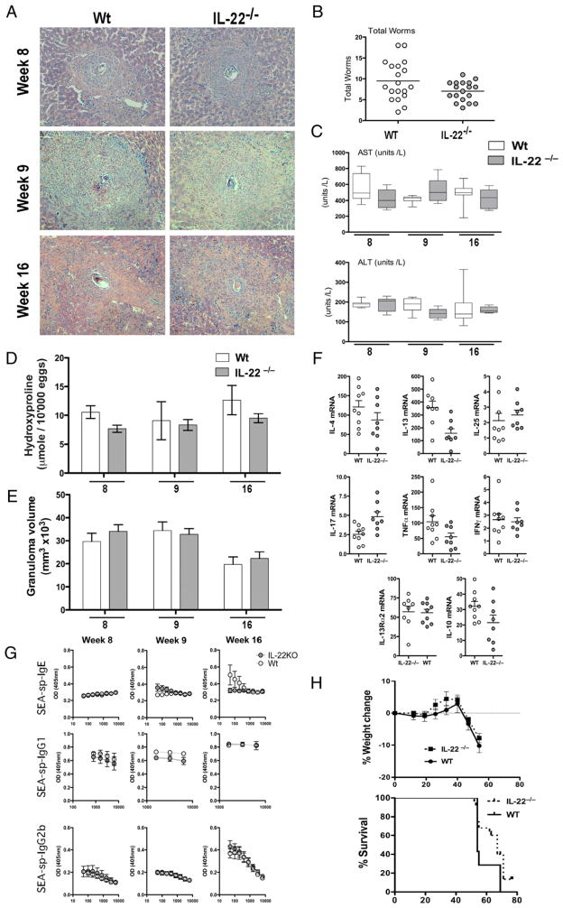
FIGURE 2.
Th2 development during acute and chronic infection with S. mansoni is unaffected in the absence of IL-22. Mice were infected with 35 cercariae, unless indicated, with worm burden, hepatic pathology, and immunological parameters assessed at week 8, 9, or 16 postinfection. One of three experiments with mean ± SEM (C–E, G, H), or individual mice per data point (B, F) are shown. A, Giemsa-stained liver section taken during acute (week 8), peak (week 9), and chronic (week 16) phases of S. mansoni infection (×10 magnification). B, Susceptibility and infection intensity expressed as worm burden. C, Serum AST and ALT levels throughout infection. D, Liver fibrosis expressed as hydroxyproline content per 10,000 eggs throughout infection. E, Granuloma volume determined from histological analysis. F, mRNA for IL-4, IL-13, IL-25, IL-17A, TNF-α, IFN-γ, IL-13Rα2, and IL-10 at week 9 of infection. G, Serum Ab isotypes specific for SEAs throughout infection. H, Weight change and percentage Kaplan–Meier graph showing survival following a high-dose (100 cercariae) infection.

