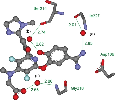
Fig. 1.
The crystal structure of Factor Xa bound with the inhibitor ZK-807834 from PDB ID 1FJS. The inhibitor is displayed as atom-colored balls and sticks and the three water molecules are displayed as red balls and labeled (a), (b) and (c) in black. Important interactions are marked in green, distances are given in Å, and protein residues Asp189, Ser214, Gly218 and Ile227 are displayed as atom colored sticks and named in green. Some atoms are not shown, for clarity.

