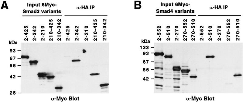
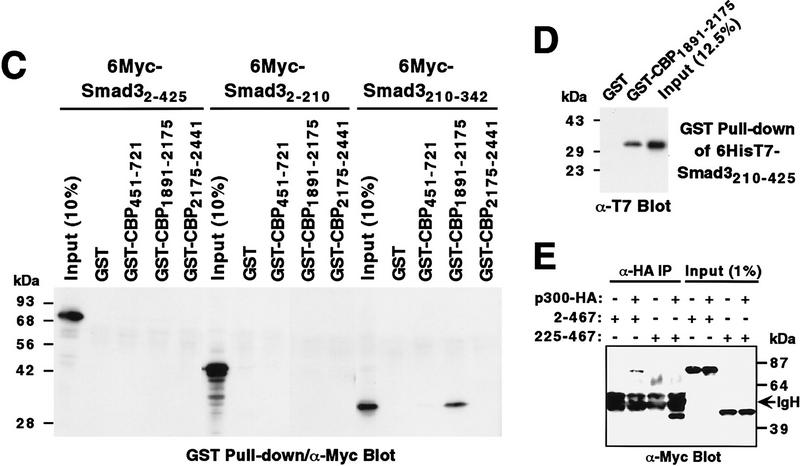
Figure 2.
Mapping of interaction domains. α-HA coimmunoprecipitations of Myc-tagged Smad3 (A) or Smad4 (B) proteins with HA-tagged p300. (Input) Approximately 2% of Myc-tagged proteins in the cell extracts. Numbers at top refer to amino acids. (C) GST pull-down assays. Bound Myc-tagged Smad proteins were revealed by α-Myc Western blotting. (D) GST pull-down assay with 6HisT7–Smad3210–425 followed by α-T7-tag Western blotting. (E) α-HA coimmunoprecipitations of Myc-tagged Smad2 proteins with p300–HA.


