Figure 3.
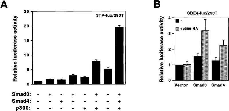
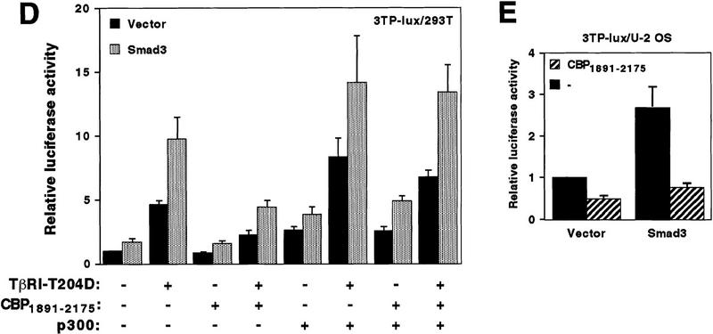
Coactivation mediated by p300 and Smad proteins. (A) 6Myc–Smad32–425 (0.1 μg), 6Myc–Smad42–552 (0.3 μg), and p300–HA (1 μg) were cotransfected, with the 3TP–lux reporter construct into 293T cells. Relative luciferase levels are depicted. (B) One microgram of 6Myc–Smad32–425, 6Myc–Smad42–552, or the empty expression vector was cotransfected with or without 0.7 μg of p300–HA into 293T cells. Relative luciferase activities derived from the cotransfected SBE4–luc reporter are presented. (C) Mv1Lu cells were transfected with the 3TP–lux reporter construct and additionally with 3 μg of p300–HA, 0.1 μg of Myc–Smad3, or 0.6 μg of Smad4–HA expression vectors. Relative luciferase activities are given. (D) Twenty-five nanograms of Myc–Smad3 expression plasmid or the empty vector pcDNA3 was cotransfected with combinations of 40 ng of TβRI–T204D, 1 μg of pEV3S–CBP1891–2175, and 0.1 μg of p300–HA plasmids into 293T cells. Luciferase activity derived from the cotransfected 3TP–lux reporter construct is depicted. (E) U2 OS cells were transfected with 100 ng of Myc–Smad3 plasmid, or pcDNA3, and the 3TP–lux reporter gene construct. Where indicated, 6 μg of pEV3S–CBP1891–2175 was cotransfected.


