
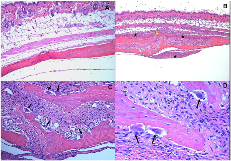
Figure 4.
Effects of polymicrobial infections with P. gingivalis/T. denticola/T. forsythia on mouse calvaria. Live P. gingivalis+T. denticola+T. forsythia (5 × 10⁸ cells/each) bacteria as polymicrobial consortium were injected once daily for 3 days into the s.c. tissues overlying the calvaria of mice. All photomicrographs are of slides stained with hematoxylin and eosin. A. Lack of edema and inflammation in the calvarial soft tissue of the sham-infected control mouse (magnification × 10). B. A section from a polymicrobial infected animal demonstrates significant disruption of the suture area (yellow arrow) with an intense mixed inflammatory infiltrate consisting primarily of neutrophils, lymphocytes and macrophages along with areas of edema and increased vascularity (magnification × 5). Inflammation is noted on both the dermal and supraosteal areas (asterix).C. Numerous osteoclasts (black arrows) are seen throughout the inner aspects of the calvarial bone mainly in the suture area. (magnification × 20). D. Activated osteoclasts within resorption lacunae at higher magnification (× 40).