
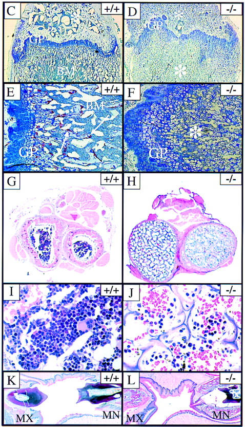
Figure 2.

Osteopetrosis in RANK−/− mice. (A,B) Radiograph of 6-week-old RANK+/+ (left) and RANK−/− (right) mice. Note the absence of the bone marrow cavity, shortening of the long bones, and overall increase in radiodensity in the RANK−/− animals. (C–L) Histological analysis of bone. (C,D) Histological staining of the femur (4× magnification). The growth plate (GP) is distorted in the RANK−/− animals and the bone marrow (BM) is filled with cartilage (*). (E,F) Histological staining of the femur (20× magnification). TRAP positive (red) osteoclasts (arrows) are readily seen in the RANK+/+ bone. Note the absence of TRAP-positive osteoclasts in RANK−/− bone and the disordering of the chondrocytes at the growth plate. (G,H) Cross section of radius and ulna indicating the occlusion of the bone marrow space in RANK−/− mice. Note the thinning of the cortical bone in RANK−/− mice. (I,J) Histological sections of vertebral bone. Note osteopetrotic condition of RANK−/− vertebrae and residual, small hematopoietic foci in the RANK−/− animal. (K,L) Histological section of the jaw, illustrating the maxilla (MX) and mandible (MN). The teeth are erupting through the wild-type jaw; in the RANK−/− mice, teeth are present but impacted in abnormal bone.