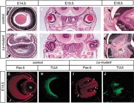
Figure 5.
Several neuroretinas form in the absence of all lens structures in the Le-mutant mice. Hematoxilin-eosin (HE) staining (A–F) of transverse (B,C,E,F) or sagittal sections (A,D) through the eye region of the Le-mutant (D–F) and control littermates; Paxflox/Pax6lacZ (A,C) or Pax6+/Pax6+ (B). Cell layers including nerve fiber layer (nfl), inner nuclear layer (inl), and outer nuclear layer (onl) are distinguished in the Le-mutant (F) and in the control eye (C) at E18.5. The black arrows point out the RPE (B–F). Note that the axons exit each retina fold not from the optic disc but, rather, from an opening between the folds, which in a normal eye would have been occupied by the lens (white arrowheads in E,F). Furthermore, an optic nerve is observed in Le-mutant eyes (F). Coimmunolabeling with antibodies to Pax6 (G,I) and to βIII tubulin (H,J) show that each retina fold develops autonomously in the Le-mutant eye. Enhanced Pax6 expression is marked with arrowheads, RPE with two arrowheads. (le) Lens; (onh) optic nerve head; (*) unspecific fluorescence from blood cells.

