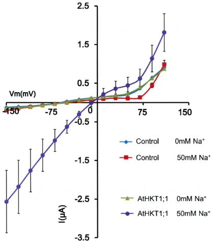
Figure 4. Removal of extracellular Na+ in AtHKT1;1 expressing Xenopus oocytes reduces AtHKT1;1-mediated outward currents.
Recordings were carried out 1 to 2 days after AtHKT1;1 cRNA injection (source: Columbia ecotype). Oocytes were clamped in perfusion solutions containing either 0 mM NaCl or 50 mM NaCl. Applied membrane potentials ranged from +110 to −150 mV. Mean steady-state currents (±SEM) recorded in oocytes injected with either 20 ng of Col-0 wild-type AtHKT1;1 (n = 8) cRNA, or with H2O (n = 13) as control.

