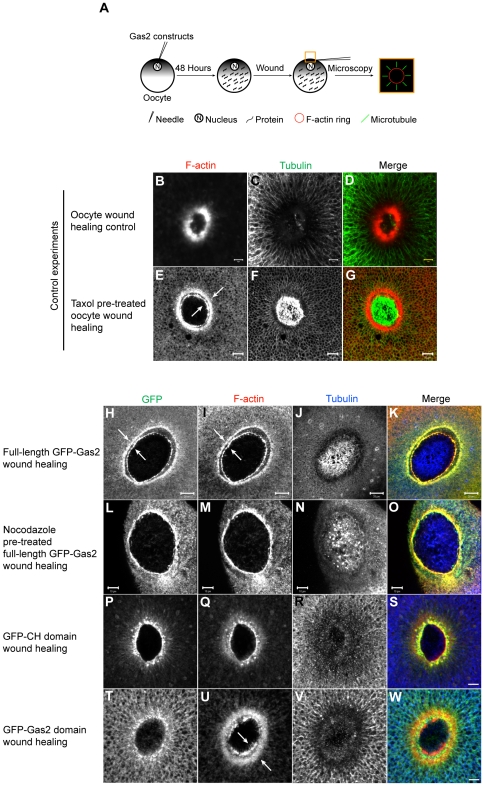
Figure 4. The expression of either full-length Gas2 protein or the Gas2 domain alone results in abnormal double actin rings at the wound border.
(A) The experimental flowchart and schematically represented results. Oocytes nuclei were injected with different GFP Gas2 constructs and incubated for 48 hours to allow for protein expression. The animal pole of an oocyte was then wounded, fixed and stained for F-actin and tubulin. The wound site was excised and examined by confocal microscopy. (B–D) Oocyte wound healing control experiment. F-actin forms a single ring and microtubules radially distribute around the wound. Bars, 5 µm. (E–G) Oocytes pre-treated with Taxol form abnormal double actin rings (arrows in E) during wound healing. Bars, 10 µm. (H–K) Oocytes expressing the full-length GFP-Gas2 form double Gas2 rings (arrows in H), which co-localize with double actin rings (arrows in I) during wound healing. Bars, 20 µm. (L–O) Oocytes expressing the full-length GFP-Gas2 and pre-treated with nocodazole form a single Gas2 ring, which co-localizes with single actin ring during wound healing. Bars, 10 µm. (P-S) Oocytes expressing GFP-CH domain alone form a single actin ring during wound healing. Bar, 10 µm. (T-W) Oocytes expressing GFP-Gas2 domain alone form a single Gas2 domain ring, which localizes between the double actin rings (arrows in U) during wound healing. The GFP-Gas2 domain does not co-localize with either actin ring. Bar, 10 µm.

