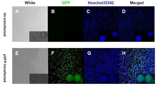
Fig. 2. Confocal imaging of GFP expression in primary human CD8+ cells.
Two million CD8+ cells were transfected with GFP plasmid or transfection buffer. After removal of dead cells, the nuclei of CD8+ cells were stained with Hoechst33342 (2 μg/ml) and cells were fixed with 4% paraformaldehyde (PFA). Confocal photos were taken using 20x and 40xZoom4x (low right corner) lens. A-D, primary human CD8+ cells were transfected with buffer as control. E-H, primary human CD8+ cells were transfected with GFP plasmid under 20ms/Pulse1/2200V conditions.

