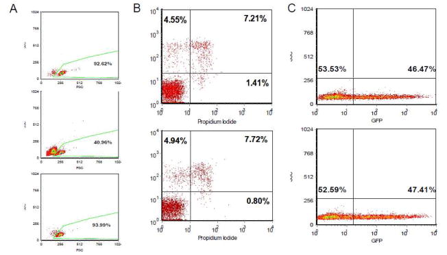
Fig. 3. Removal of Dead and dying cells.
Two million primary CD8+ cells were either untreated or transfected in 100 μl tips in Buffer T with 10 μl of 1 μg/μl GFP plasmid under the following conditions: 20ms/Pulse1/2200V. The dying and dead cells were removed using a dead cell removal kit. A) Cell viability was tested by flow cytometry (BD FACsort) 24 hours after electroporation. Upper panel: untreated cells; middle panel: cells prior to dead cell removal; less panel: cells after dead cell removal. B) Untreated primary CD8+ cells (upper panel) and transfected CD8+ cells after dead cell removal (less panel). The cells were stained with Annexin and Propidium Iodide. C) The transfection efficiency was not changed by dead cell removal. The percentage of GFP-positive cells was tested before (upper panel) and after (less panel) dead cell removal.

