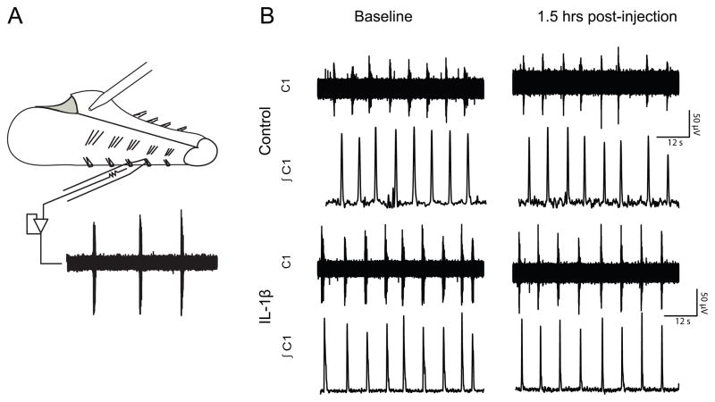
Figure 3.
Fictive motor output was recorded from cranial nerve rootlets. (A.) An illustration representing the in vitro en bloc brainstem-spinal cord preparation. Injections (2μL) were made in the caudal, commisural NTS and motor output was recorded from cranial nerve rootlets using glass suction electrodes. (B) A sample trace from a control preparation receiving saline and a preparation receiving an IL-1β (10μg) injection. Raw data (top traces) were integrated (bottom traces) and analyzed to determine changes in breathing patterns. The traces are 1 min segments.

